Diagnostic ultrasound is a non-invasive imaging test that uses high-frequency sound waves to produce live images of internal structures. It does not use radiation. Physicians order it to investigate symptoms, confirm or rule out conditions, and guide clinical decisions across a wide range of specialties.
What Is Diagnostic Ultrasound
Diagnostic ultrasound is a medical imaging method that transmits sound waves into the body at frequencies far above the range of human hearing. According to Health Canada’s medical ultrasound overview, frequencies used in diagnostic imaging range 100 to 500 times higher than what the human ear can detect. When those waves contact internal structures, they reflect back as echoes. A transducer on the skin captures the returning signals, which are processed into live images on a monitor.
The technique produces still images and moving sequences, depending on what is being assessed. It is safe for use in all patient populations, including pregnant patients, because it does not involve ionizing radiation. Scans are performed in accredited diagnostic imaging facilities by registered sonographers, and the resulting images are reviewed and reported by a radiologist.
What Diagnostic Ultrasound Shows in Clinical Practice

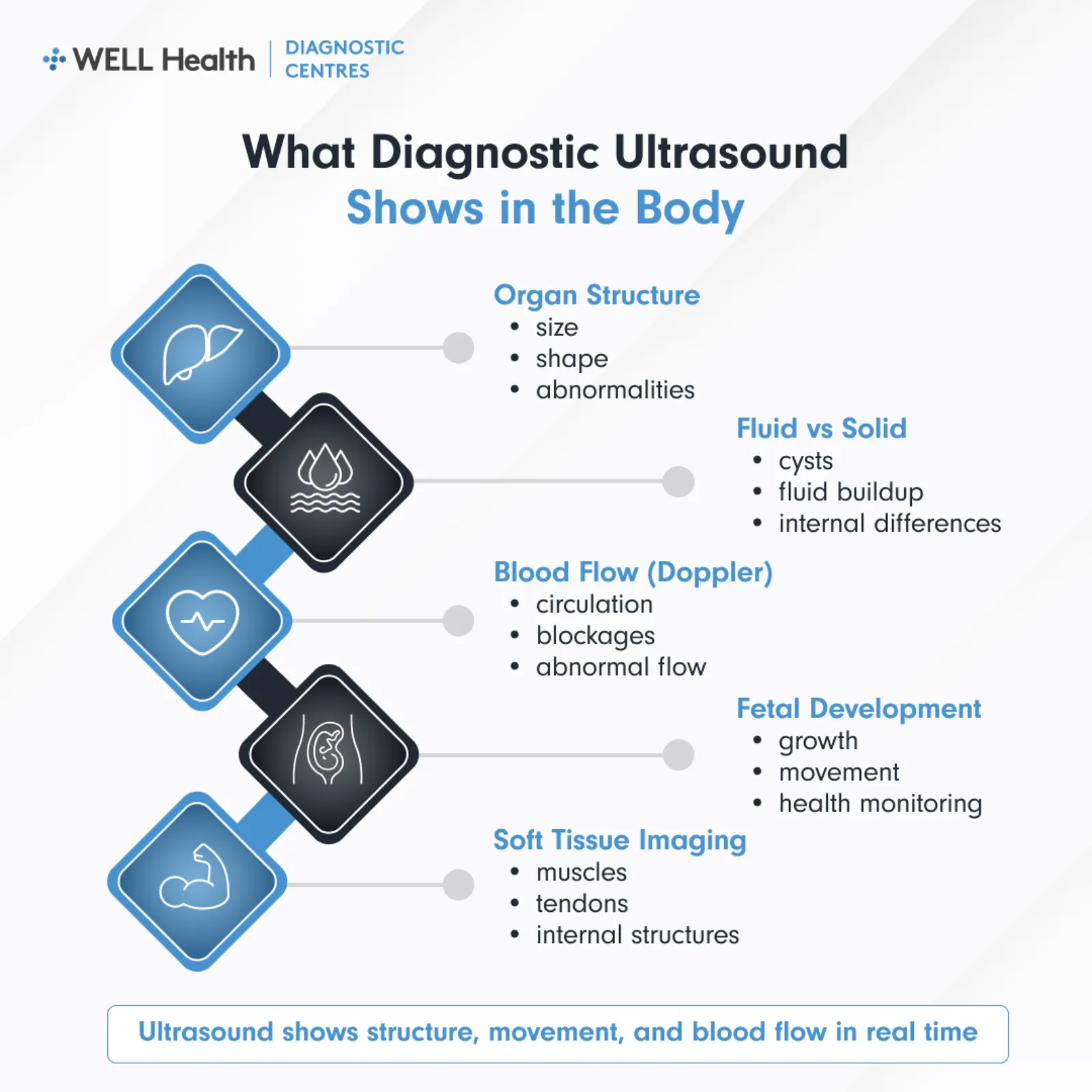
The Ontario Association of Radiologists’ patient information on ultrasound notes that the test captures moving images of pelvic and abdominal structures, and that a Doppler enhancement allows it to document blood flow in vessels and cardiac movement. What the scan shows depends on the body area being examined and the clinical question the physician is trying to answer.
Organ Structure and Size
Ultrasound shows the size, shape, and internal texture of solid organs. This includes the liver, kidneys, gallbladder, pancreas, spleen, thyroid, and bladder. Physicians use these images to identify enlargement, structural irregularities, cysts, and masses that warrant further investigation.
Fluid and Soft Tissue
The test distinguishes between solid tissue and fluid-filled structures. It identifies free fluid in the abdomen or pelvis, fluid collections around organs, and the internal characteristics of cysts. This distinction informs if a structure is likely benign or requires follow-up imaging or biopsy.
Blood Flow and Vascular Structures
Doppler ultrasound measures blood flow velocity and direction in arteries and veins. It is used to assess circulation in the carotid arteries, renal arteries, deep leg veins, and portal system. The results help identify narrowing, blockages, or abnormal flow patterns associated with vascular conditions.
Fetal Development
Obstetric ultrasound produces images of the fetus during pregnancy. The Health Canada overview states that fetal ultrasound provides information on the size, age, and health of the fetus, and can detect multiple pregnancies and certain birth defects. Growth scans at various stages of pregnancy track fetal development against established clinical benchmarks.
When Diagnostic Ultrasound Is Ordered
Physicians order a diagnostic ultrasound when a patient presents with symptoms that require imaging to assess internal structures. It is also ordered as part of routine monitoring, follow-up after previous findings, and obstetric care. In Ontario, the scan requires a signed requisition from a licensed healthcare provider.
- Abdominal pain or discomfort: To assess the liver, gallbladder, kidneys, pancreas, spleen, and surrounding structures.
- Pelvic symptoms: To evaluate the uterus, ovaries, and bladder in patients reporting pelvic pain, abnormal bleeding, or urinary symptoms.
- Suspected kidney stones: To identify obstructions or calcifications in the renal system.
- Thyroid abnormalities: To characterize nodules identified on physical examination or other imaging.
- Vascular assessment: To evaluate blood flow in the carotid arteries, lower limb veins, or abdominal vessels.
- Obstetric monitoring: To assess fetal growth, position, placenta, and amniotic fluid at scheduled intervals.
- Scrotal concerns: To examine the testes and surrounding structures for masses, inflammation, or torsion.
- Musculoskeletal symptoms: To assess soft tissue, tendons, and joints when X-ray findings are inconclusive.
Common Conditions Evaluated Using Ultrasound
The General Ultrasound page on RadiologyInfo.org, a resource produced by the Radiological Society of North America, outlines the range of conditions that ultrasound imaging is used to evaluate in clinical practice. The following represent the most frequently assessed categories.
Gallbladder and Biliary System
Gallstones are among the most common findings on abdominal ultrasound. The scan also assesses bile duct dilation, gallbladder wall thickening, and signs of acute cholecystitis. These findings directly inform decisions about surgical referral and further investigation.
Liver and Kidneys
Ultrasound evaluates liver texture for signs of fatty change, cirrhosis, or focal lesions. For the kidneys, it assesses size, cortical thickness, cysts, hydronephrosis, and the presence of stones. It is a first-line imaging test for both organs before more advanced imaging is considered.
Pelvic and Reproductive Structures
In female patients, ultrasound assesses the uterus and ovaries for fibroids, cysts, masses, and structural changes. In male patients, scrotal ultrasound examines the testes for torsion, masses, or varicoceles. Pelvic ultrasound is also the primary imaging method in obstetric care.
Deep Vein Thrombosis and Vascular Conditions
Venous ultrasound of the lower limbs is used to rule out or confirm deep vein thrombosis. Carotid ultrasound assesses plaque buildup and arterial narrowing associated with stroke risk. Both are ordered based on specific clinical presentations and risk factors.
What to Expect During the Procedure
The Ontario Association of Radiologists describes ultrasound as a simple, safe, and painless procedure. The scan involves no injections and no radiation exposure of any kind. Most appointments are completed within 20 to 45 minutes, depending on the area being examined.
- Positioning: You will lie on a padded table, positioned to allow access to the area being scanned.
- Gel application: A clear water-based gel is applied to the skin over the scan area to improve sound wave transmission.
- Transducer use: The sonographer moves a handheld transducer across the skin to capture images.
- Real-time imaging: Images appear on a monitor during the scan and are recorded for radiologist review.
- Post-scan: The gel wipes off easily. No recovery period is required and you can resume normal activities immediately.
For internal structures such as pelvic organs, a transvaginal transducer may be used with the patient’s consent. The sonographer will explain the procedure before it begins.
How Results Are Interpreted and Reported
A radiologist reviews the ultrasound images and prepares a written diagnostic report. That report is transmitted to your referring healthcare provider, who then contacts you to discuss the findings. The radiologist does not communicate results directly to patients.
Well Diagnostics transmits diagnostic reports electronically to referring providers. Patients can also access their images and reports through PocketHealth, a secure platform that allows you to view, download, and share your imaging records once the report is finalized.
Key Takeaways
- No radiation exposure: Diagnostic ultrasound uses sound waves, not X-rays or ionizing radiation.
- Wide clinical application: It evaluates abdominal organs, pelvic structures, blood vessels, soft tissue, and fetal development.
- Ordered by referral: A signed requisition from a licensed healthcare provider is required in Ontario.
- Radiologist interprets findings: A written report is sent to your referring physician, who reviews it with you.
- Requires no recovery time: Patients return to normal activities immediately after the scan.
Frequently Asked Questions
What does a diagnostic ultrasound show that other tests cannot?
Ultrasound shows real-time soft tissue images and fluid movement that standard X-rays cannot produce. It also captures blood flow through Doppler imaging, which is not available through static imaging methods. For soft tissue structures, fluid-filled spaces, and fetal assessment, it is often the first and most appropriate imaging test ordered.
Is diagnostic ultrasound the same as an ultrasound imaging test?
Yes. The terms refer to the same procedure. Diagnostic ultrasound and ultrasound imaging tests both describe the use of high-frequency sound waves to produce internal images for clinical assessment. The word diagnostic is used to distinguish it from therapeutic ultrasound, which is a separate application used in physiotherapy and surgical procedures.
Do I need a referral for a diagnostic ultrasound in Ontario?
Yes. In Ontario, a signed requisition from a licensed healthcare provider is required for an OHIP-covered diagnostic ultrasound. If you do not have a referring physician, you can obtain a requisition from a family doctor, walk-in clinic, or community health centre. Well Diagnostics accepts requisitions from any certified Ontario healthcare facility.
Diagnostic Ultrasound as a Clinical Assessment Tool
Diagnostic ultrasound provides organ structure, fluid assessment, vascular flow, and fetal imaging without radiation exposure. Physicians order it when symptoms point to conditions in the abdomen, pelvis, vessels, or soft tissue that require visual confirmation. Results are reviewed by a radiologist and communicated to your referring provider, who discusses findings and next steps with you.