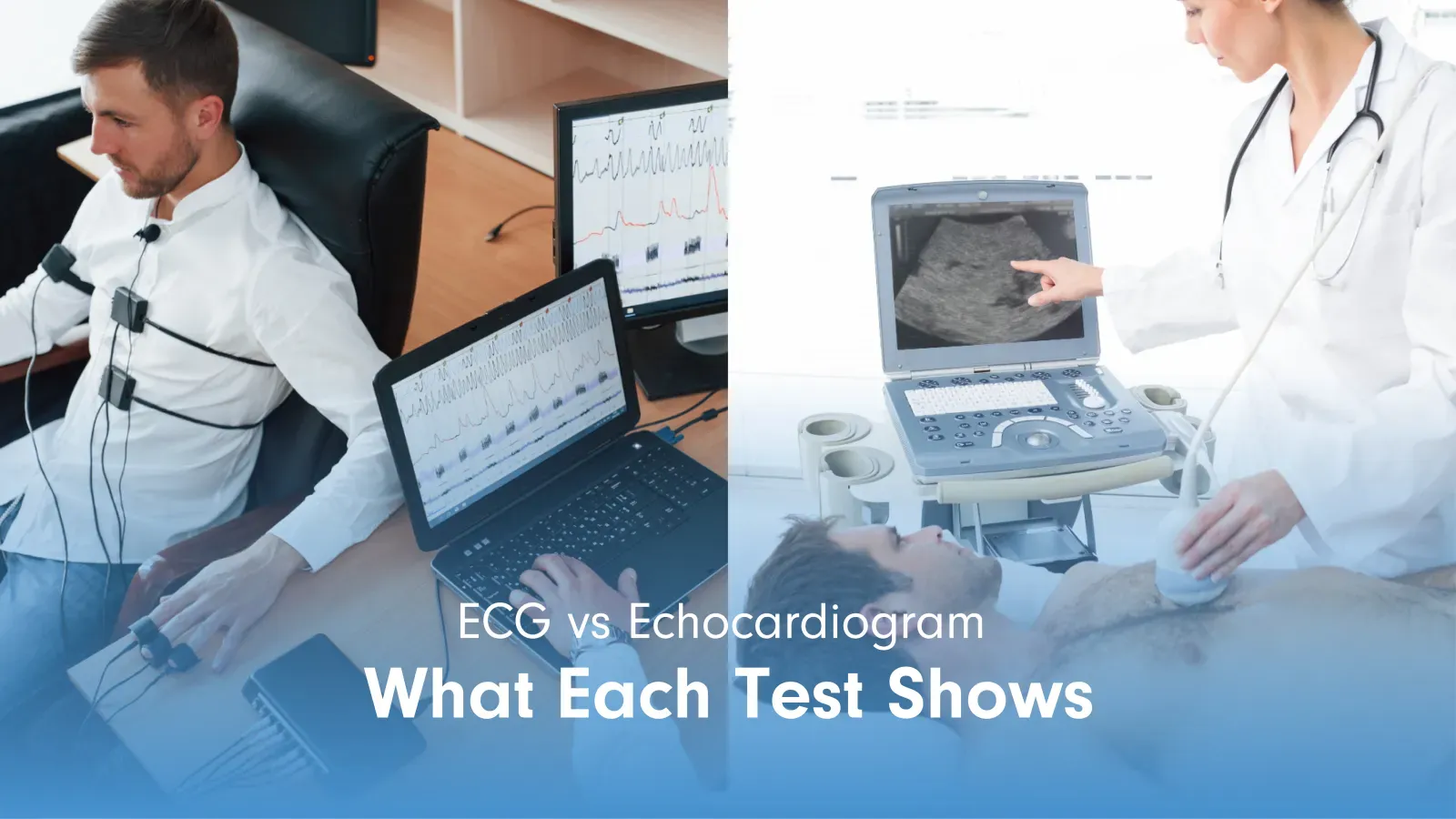
Cardiac diagnostic testing covers a range of tests, each measuring a different aspect of heart function. No single test provides a complete picture. Physicians select the appropriate test based on the patient’s symptoms, risk factors, and previous test results.
Overview of Cardiac Tests
Cardiac tests help doctors understand how the heart is working, why symptoms may be happening, and if there are signs of heart disease. These tests do not all look for the same thing; some focus on electrical activity, some create images of the heart’s structure, and others show how well the heart performs during activity or over time.
In general, cardiac diagnostic tests are grouped into three broad categories:
- Electrical tests: Record the heart’s rhythm, rate, and electrical signals to help detect issues such as irregular heartbeats or conduction problems.
- Imaging tests: Create pictures of the heart, valves, chambers, and blood vessels to help evaluate structure and movement.
- Functional tests: Assess how the heart performs during stress, activity, or extended monitoring when symptoms may come and go.
Together, these tests give doctors different pieces of information. An ECG may show how the heart’s electrical system is behaving, while an echocardiogram may show how the heart’s structure and pumping function look.
When Cardiac Tests Are Ordered
The clinical presentation determines which test is selected first and which follow-up tests may be needed.
- Palpitations or irregular rhythm: ECG first; Holter monitor if ECG is normal, but symptoms persist.
- Chest pain at rest: ECG immediately; troponin blood test to assess for myocardial damage.
- Chest pain on exertion: Exercise stress test or nuclear stress test to assess coronary perfusion under load.
- Shortness of breath: Echocardiogram to assess ventricular function and valve status.
- Heart murmur: Echocardiogram to characterize valve abnormality and assess severity.
- Fainting or near-fainting: ECG and Holter monitor to rule out electrical causes; echocardiogram if structural cause suspected.
- Pre-surgical cardiac clearance: ECG at minimum; echocardiogram if significant cardiac history or elevated surgical risk.
What Cardiac Tests Show in Diagnosis
Different heart tests tell doctors different things about how the heart is working. Some show if the heartbeat is steady, some show how the heart looks, and pumps, and others show how it responds to activity or stress.
Looking at these results together helps doctors better understand what may be causing symptoms and what kind of care may be needed next.
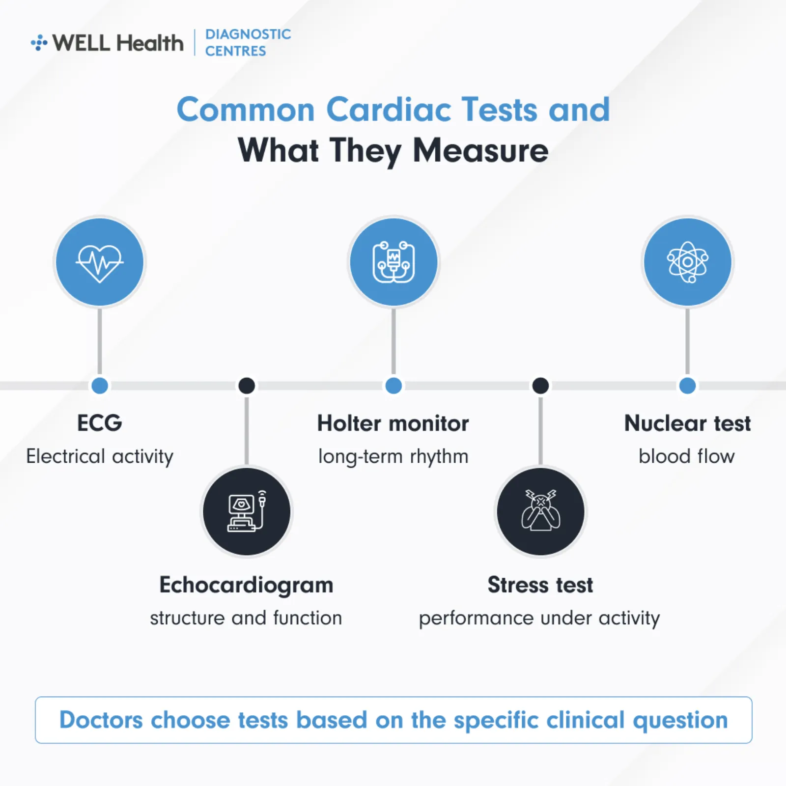
Electrocardiogram (ECG)
An ECG records the electrical activity of the heart using electrodes placed on the chest, arms, and legs. It shows heart rate, rhythm, and the pattern of electrical conduction through the heart muscle. Physicians use ECG findings to identify arrhythmias, evidence of prior heart attack, conduction delays, and chamber enlargement.
Echocardiogram
An echocardiogram uses ultrasound to produce real-time images of the heart’s four chambers, valves, walls, and major vessels. It measures ejection fraction, assesses valve function, identifies wall motion abnormalities, and detects structural defects. It is the primary imaging test for evaluating heart failure, valve disease, and cardiomyopathy.
Holter Monitor
A Holter monitor is a portable ECG device worn for 24 to 48 hours. It records continuous heart rhythm data during normal daily activities and sleep. Physicians order it when intermittent arrhythmias are suspected but not captured on a standard ECG.
Exercise Stress Test
An exercise stress test records ECG and blood pressure data while the patient walks on a treadmill or cycles under a gradually increasing load. It assesses how the heart responds to physical demand and helps identify exercise-induced arrhythmias or ischaemia. It is ordered when symptoms such as chest pain or shortness of breath occur, specifically during exertion.
Nuclear Stress Test
A nuclear stress test combines exercise or pharmacological stress with a radiotracer injection to image blood flow through the heart muscle. It identifies areas of reduced perfusion that indicate coronary artery disease or prior infarction. It is ordered when an exercise stress test result is inconclusive or when a patient cannot exercise adequately.
Common Types of Cardiac Testing
Cardiac tests are not one-size-fits-all. Doctors choose different tests depending on what they need to check, such as the heart’s rhythm, structure, blood flow, or response to activity. The most common tests usually fall into a few main groups, each giving a different kind of information about heart health.
Electrical Activity Tests
The ECG and Holter monitor both measure electrical signals from the heart, differing only in duration. An event recorder extends monitoring beyond 48 hours for arrhythmias that occur very infrequently. Implantable loop recorders are used in rare cases where arrhythmias are suspected but not captured even after prolonged external monitoring.
Cardiac Imaging Tests
Echocardiography is the most widely ordered cardiac imaging test in outpatient practice. A transesophageal echocardiogram (TEE) places the ultrasound probe behind the heart via the esophagus for higher-resolution images of posterior structures. Cardiac MRI and CT are available for specific clinical questions, including detailed tissue characterization and coronary artery assessment.
Functional and Perfusion Tests
Stress testing, if the exercise-based or pharmacological, assesses coronary perfusion and exercise tolerance. Nuclear cardiology adds perfusion imaging to identify which areas of the heart muscle are receiving adequate blood flow. These tests are most often ordered for patients with suspected coronary artery disease or known risk factors.
How Results Guide Clinical Decisions
Cardiac test results help doctors move from a broad concern to a more specific understanding of what may be happening with the heart. A result may confirm a suspected diagnosis, rule out certain problems, or show that another test is needed to get a clearer picture.
- Confirming a diagnosis: Test results can support a diagnosis such as an arrhythmia, valve disease, heart failure, coronary artery disease, or a prior heart attack.
- Deciding if more testing is needed: A normal result does not always end the evaluation, especially if symptoms continue or occur only during certain activities.
- Matching the test to the symptom: For example, palpitations that do not appear on a standard ECG may lead to a Holter monitor or longer rhythm monitoring.
- Assessing severity: Results can show if a condition is mild, moderate, or severe, which helps guide treatment urgency and follow-up.
- Planning treatment: Findings such as reduced ejection fraction, abnormal valve function, or evidence of reduced blood flow may lead to medication changes, lifestyle guidance, or referral to a cardiologist.
- Considering the full clinical picture: Doctors interpret results alongside symptoms, medical history, physical exam findings, and risk factors rather than relying on one test alone.
Next Steps After Cardiac Testing
After testing is complete, your cardiologist or referring physician reviews the report and explains what the findings mean in the context of your symptoms and health history. The next step depends on if the results are normal, unclear, or show a possible heart-related condition.
- Reviewing the results: Your doctor will explain if the test was normal, abnormal, or inconclusive and what that means for your care.
- Starting or adjusting medication: Some findings may lead to new medication or changes to current treatment, especially for rhythm problems, blood pressure, heart failure, or reduced blood flow.
- Recommending lifestyle changes: Your doctor may suggest changes related to exercise, diet, smoking, weight management, stress, or other risk factors.
- Ordering further testing: Additional imaging, stress testing, rhythm monitoring, or blood work may be recommended if more information is needed.
- Referring to a specialist: Abnormal or complex results may lead to referral to a cardiologist, electrophysiologist, heart failure specialist, or valve specialist.
- Setting a monitoring plan: If results are normal or stable, your doctor may recommend routine follow-up, repeat testing at a later date, or symptom-based monitoring.
Key Takeaways
- Different tests, different questions: Each cardiac test is ordered to answer a specific clinical question about rhythm, structure, function, or perfusion.
- ECG is typically the first test: Most cardiac assessments begin with an ECG because it is fast, non-invasive, and available in most clinical settings.
- Echocardiogram shows structure: When the concern involves valves, chambers, or pumping function, echocardiography is the primary imaging test ordered.
- Holter extends ECG recording: For intermittent rhythm symptoms, a 24 to 48-hour Holter monitor captures what a brief ECG cannot.
- Stress testing adds functional context: Exercise or pharmacological stress tests show how the heart performs under physiological demand.
Frequently Asked Questions
What is the most common first cardiac test ordered?
An ECG is typically the first cardiac test ordered in most clinical presentations. It is fast, widely available, non-invasive, and provides immediate information about rhythm and conduction. It serves as the baseline from which other cardiac tests are selected based on what the ECG shows or fails to show.
Can one cardiac test replace another?
No. Each cardiac test is designed to answer a different clinical question and cannot substitute for another. An ECG cannot show valve function; an echocardiogram cannot assess coronary perfusion; a stress test cannot detect structural abnormalities. Physicians use a combination of tests to build a complete assessment.
How long does a full cardiac diagnostic workup take?
The timeline depends on the tests ordered and the clinical urgency. An ECG is completed in under 10 minutes. An echocardiogram takes 30 to 60 minutes. A Holter monitor requires 24 to 48 hours of wearing the device. Results from each test are sent to your physician within a few business days of completion.
Cardiac Tests as a Structured Assessment Pathway
Cardiac diagnostic testing follows a logical sequence based on the clinical question at each stage. Tests are not ordered at random; each one builds on the previous result to narrow the clinical picture. Your physician and cardiology team use the findings together to determine what your heart is doing and what, if anything, needs to change.