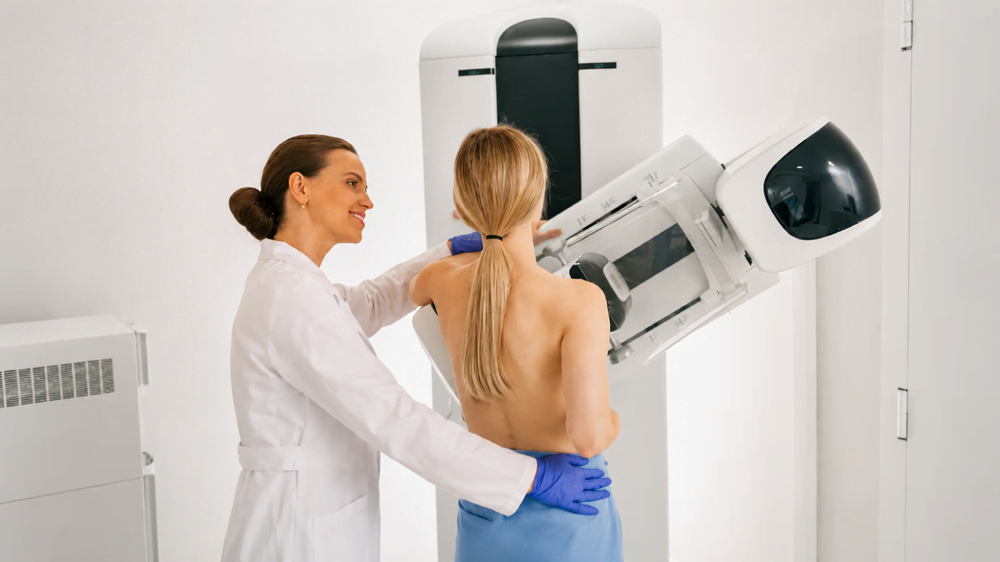
A breast ultrasound after mammogram is not a repeat of the same test. It is a different imaging method that answers a different clinical question. Physicians order it to clarify specific findings that mammography alone cannot fully characterize.
Role of Ultrasound in Breast Imaging
Ultrasound and mammography are complementary imaging tools. The Ontario Association of Radiologists’ patient resource on ultrasound describes ultrasound as a diagnostic procedure that captures real-time moving images of internal structures using high-frequency sound waves, without radiation exposure. In breast imaging, it is used alongside mammography to characterize areas that appear abnormal or ambiguous on X-ray.
The two tests show different aspects of breast tissue. Mammography is better for detecting calcifications and structural changes across the full breast. Ultrasound is better for evaluating a specific area and distinguishing solid from fluid-filled tissue.
Breast Ultrasound After Mammogram: When It Is Required

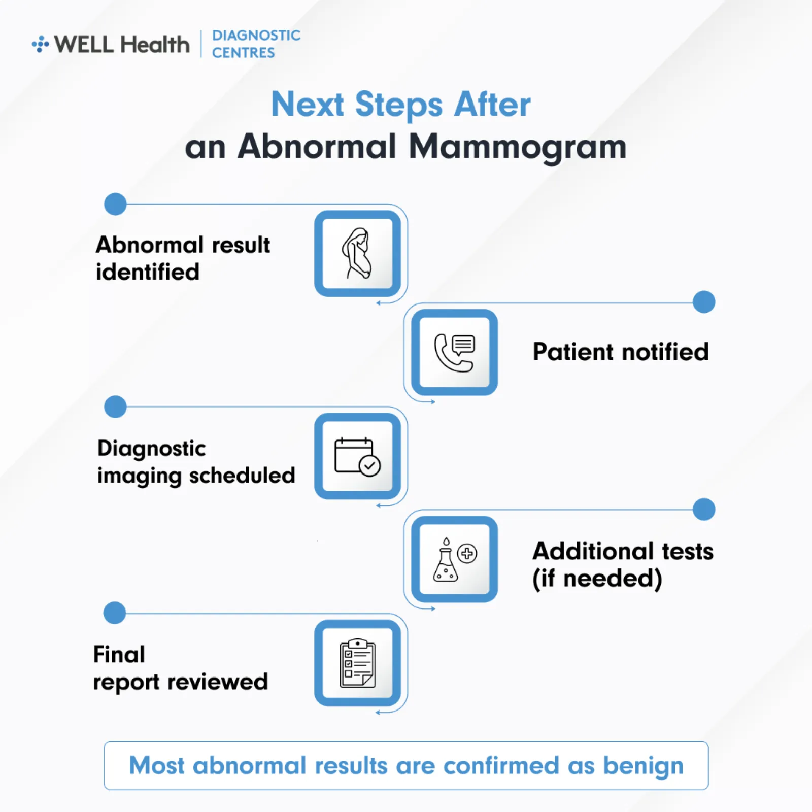
Your physician or radiologist may order a breast ultrasound after mammography in several clinical scenarios. The decision is based on the mammogram findings, your symptoms, breast density, and age.
- Abnormal screening result: When a mammogram flags an area as incomplete (BI-RADS 0), ultrasound is often the first follow-up test ordered.
- Dense breast tissue: In patients with dense breasts, mammography has reduced sensitivity; ultrasound provides supplemental tissue assessment.
- Palpable lump: If a lump can be felt but is not clearly visible on mammogram, ultrasound is used to image it directly.
- Suspected cyst: Ultrasound confirms even if a round mammographic density is a simple fluid-filled cyst or requires further investigation.
- Younger patients: For patients under 30 presenting with a breast concern, ultrasound is generally preferred over mammogram as a first-line imaging test.
What Ultrasound Can Clarify After Mammography
RadiologyInfo.org’s guidance on breast ultrasound outlines the clinical questions this test is designed to answer.
Cyst vs. Solid Mass
A simple cyst appears as a round, fluid-filled structure with smooth walls and no internal echoes on ultrasound. Simple cysts are benign and require no further investigation. A solid mass, by contrast, has internal echoes and characteristics that the radiologist assesses for further follow-up.
Characterizing a Mammographic Finding
When a mammogram shows a density, distortion, or asymmetry, ultrasound is used to determine if it corresponds to a legitimate tissue or is an artifact of overlapping structures. This step prevents unnecessary callbacks and provides a more complete picture before any biopsy decision is made. The radiologist correlates both images when preparing the final report.
Guiding Biopsy Procedures
When a breast ultrasound identifies a solid mass or suspicious area, it can also guide a core needle biopsy immediately. The sonographer or radiologist uses the ultrasound image to position the biopsy needle precisely within the area of concern. This technique reduces the need for surgical biopsy in most cases.
Differences Between Mammogram and Ultrasound Findings
The two imaging methods interpret breast tissue differently and are read side by side when both are performed.
- Calcifications: Visible on mammogram; not reliably detected on ultrasound.
- Fluid-filled cysts: Best characterized by ultrasound; may appear as a density on mammogram without further detail.
- Solid masses: Both tests assess these, but ultrasound provides immediate shape, margin, and internal texture information.
- Dense tissue: Mammogram identifies density; ultrasound images through it to assess underlying structures.
- Vascular supply: Doppler ultrasound can assess blood flow within a mass, which contributes to clinical characterization.
What to Expect During the Ultrasound
The Ontario Association of Radiologists describes breast ultrasound as a simple, painless procedure that involves no radiation. A gel is applied to the breast and a handheld transducer is moved across the skin surface to capture images. The sonographer positions you on a padded table and may ask you to raise your arm to access specific areas.
The appointment typically takes 15 to 30 minutes. No recovery period is required, and you can resume normal activities immediately. The sonographer does not share findings with you during the appointment; the radiologist reviews the images and sends a written report to your physician.
Reporting and Next Steps
Both the mammogram and breast ultrasound findings are reviewed by a radiologist, who prepares a single integrated report. That report is sent to your referring physician or OBSP provider. Depending on the findings, your physician may return you to routine screening, recommend a short-interval follow-up, or refer you for a biopsy.
Key Takeaways
- Different technology: Mammography uses X-ray; ultrasound uses sound waves to produce live soft tissue images.
- Ordered for specific findings: Ultrasound is used to assess cysts, masses, dense tissue areas, and findings flagged on mammography.
- Identifies solid vs. fluid: The main clinical value of ultrasound is distinguishing between fluid-filled cysts and solid masses.
- Common in younger patients: For patients under 30 with breast symptoms, ultrasound is typically the first imaging test ordered.
- Results go to your physician: A radiologist reviews both the mammogram and ultrasound and sends a combined report to your referring provider.
Frequently Asked Questions
Does a breast ultrasound always follow a mammogram?
No. Ultrasound is ordered when there is a specific clinical reason, not as a routine companion to every mammogram. It is used when mammography findings require further characterization, when breast density limits mammography sensitivity, or when a patient presents with a palpable symptom. Your referring physician determines if an ultrasound is needed based on your specific imaging results.
Is breast ultrasound covered by OHIP in Ontario?
Yes, when ordered by a licensed Ontario healthcare provider through a signed requisition. Ultrasound performed as part of follow-up imaging after an abnormal mammogram or to investigate a breast symptom is OHIP-covered at accredited diagnostic imaging centres. Contact your physician or the imaging centre if you have questions about coverage for your specific appointment.
What if my breast ultrasound also shows something abnormal?
An abnormal ultrasound finding does not confirm cancer. The radiologist will characterize the finding using clinical criteria and may recommend short-interval follow-up, additional imaging, or biopsy depending on the result category. Your physician reviews the report and advises on the appropriate next step based on the combined imaging findings.
Breast Ultrasound as Part of the Imaging Pathway
Breast ultrasound after mammography is a standard and well-established step in the breast imaging pathway. It adds clinical information that mammography alone cannot provide and helps determine if a finding requires further action or confirms a benign result. Your physician and the radiologist use both sets of images together to guide what comes next.