An X-ray is a form of diagnostic imaging that uses ionizing radiation to produce two-dimensional images of internal structures. It is most commonly used for bone assessment, chest evaluation, and abdominal imaging. Physicians order X-rays when a clinical question can be answered by this imaging method, and when the benefit of the information outweighs the small radiation risk associated with the exam.
Key Takeaways
- Ordered after clinical examination: Health Canada guidance states that X-ray should not be performed as routine screening without a prior clinical evaluation.
- Most useful for bone and lungs: X-ray provides the highest diagnostic value for skeletal injuries, chest pathology, and abdominal gas patterns.
- Radiologist reports findings: Images are reviewed by a radiologist who sends a signed diagnostic report to your referring physician.
- Radiation exposure is low: The clinical benefit of a needed X-ray consistently outweighs the small radiation risk from a single diagnostic exam.
- Soft tissue requires other imaging: MRI or ultrasound is ordered when the clinical question involves tendons, ligaments, muscles, or internal organs.
Role of X-Ray in Diagnostic Imaging
X-ray is the most widely available and fastest diagnostic imaging tool in clinical practice. Health Canada’s guidance on medical and dental X-rays states that diagnostic X-ray exams provide important information about medical conditions, and that the risk of not having a needed X-ray can be much greater than the risk from radiation exposure. The decision to order an X-ray is always based on a clinical evaluation first; routine imaging without a specific indication is not recommended.
When Is an X-Ray Needed in Diagnosis
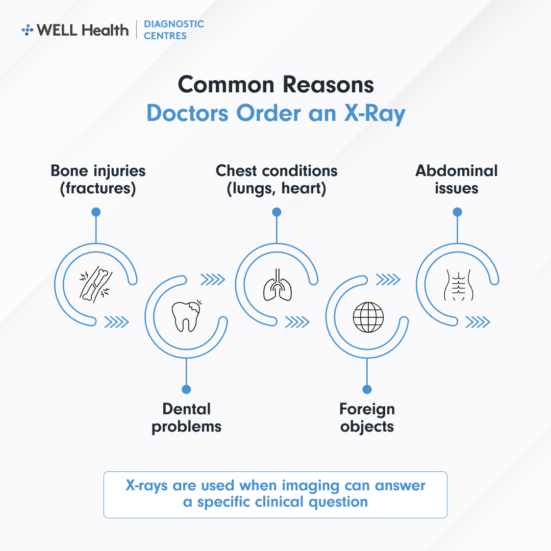
An X-ray is ordered when the suspected condition involves formations that produce clear contrast on standard radiography, primarily bone, and secondarily the lungs, heart borders, and gas-filled spaces.
Bone and Musculoskeletal Assessment
The Ontario Association of Radiologists’ patient resource on X-ray describes bone radiography as one of the most common imaging applications, used in hospitals, emergency departments, and orthopedic clinics every day. X-ray images of the skull, spine, joints, and extremities are ordered to identify fractures, including hairline and stress fractures, bone chips, joint alignment, and post-treatment positioning. Bone X-rays are also used as a baseline in orthopedic surgical planning and post-operative follow-up.
Chest and Respiratory Evaluation
A chest X-ray is one of the most commonly ordered diagnostic images. It assesses the lungs for consolidation, pleural effusion, pneumothorax, and masses; the heart for size and border clarity; and the mediastinum for widening or structural changes. It is typically the first imaging test ordered in presentations of cough, shortness of breath, chest pain, and fever with suspected respiratory infection.
Abdominal and Gastrointestinal Use
Abdominal X-ray can identify bowel obstruction through air-fluid levels and dilated loops of bowel, free air under the diaphragm indicating perforation, and calcified kidney stones or gallstones. It is used as an initial screen in acute abdominal presentations before more detailed imaging, such as CT or ultrasound, is arranged. In pediatric patients, it is also used to identify swallowed foreign objects.
Dental and Maxillofacial Applications
Dental X-rays are used to identify decay between teeth, bone loss from gum disease, impacted teeth, and changes in root structure. Health Canada states that dental X-rays should only be performed after a clinical examination to obtain diagnostic information or monitor treatment. They should not be conducted as routine screening without a clinical basis.
Conditions Commonly Evaluated Using X-Ray
- Fractures and bone injury: X-ray identifies acute fractures, healing progress, and post-treatment alignment across all skeletal regions.
- Pneumonia: Consolidation in a lung lobe produces a characteristic opacity on chest X-ray that supports a clinical diagnosis.
- Heart failure: Cardiomegaly and vascular congestion patterns visible on chest X-ray support clinical assessment of heart failure.
- Arthritis and joint disease: Joint space narrowing, erosions, and degenerative changes are visible on plain radiography.
- Bowel obstruction: Air-fluid levels and dilated bowel loops on abdominal X-ray indicate potential obstruction requiring urgent management.
- Foreign body identification: Metallic or dense objects in the body or gastrointestinal tract are visible on plain X-ray.
- Post-surgical review: X-ray confirms hardware positioning, implant placement, and fracture healing in orthopedic follow-up.
How X-Ray Findings Support Diagnosis
X-ray findings support the diagnosis by confirming or excluding specific conditions that have characteristic imaging appearances. A radiologist analyzes the images and prepares a signed report for your referring physician. The OAR notes that the radiologist may also recommend treatment in straightforward cases, such as casting for a simple fracture, or referral to a specialist when findings require further clinical management.
X-ray findings are always interpreted in the context of clinical information. A normal X-ray does not rule out every condition, and an abnormal finding does not always require treatment. Your physician combines the imaging report with your symptoms and history to determine the next step.
Limitations of X-Ray Imaging
X-ray is effective for dense structures but has well-defined limitations. Soft tissue injuries, including muscle tears and ligament damage, are not reliably visible on plain radiography and typically require MRI for assessment. Early bone stress reactions and cartilage pathology may not be apparent on X-ray before structural damage has occurred. In these situations, ultrasound, CT, or MRI is ordered depending on the specific clinical question.
When Additional Imaging Is Required
If X-ray findings are inconclusive or the clinical question involves soft tissue, vascular, or neurological formation, additional imaging is ordered. CT provides cross-sectional detail of bone and soft tissue with higher sensitivity than plain radiography. MRI provides superior soft tissue characterization without radiation exposure. Ultrasound is preferred for soft tissue, fluid, and vascular assessment when real-time imaging is needed.
Frequently Asked Questions
Do I need a referral for a diagnostic X-ray in Ontario?
Yes. In Ontario, OHIP-covered X-ray requires a signed requisition from a licensed healthcare provider. X-ray at Well Diagnostics locations is available on a walk-in basis, but a valid requisition from your physician or nurse practitioner is required at the time of the visit. If you do not have a referring provider, you can obtain a requisition from a walk-in clinic before attending the imaging appointment.
Is a diagnostic X-ray safe during pregnancy?
Physicians generally avoid X-rays of the abdominal and pelvic regions during pregnancy to minimize radiation exposure to the developing fetus. Health Canada states that when an X-ray is clinically necessary during pregnancy, technologists use specific techniques to reduce radiation dose to the lowest level consistent with obtaining a diagnostic image. Always inform your physician and the radiographer if you are pregnant or may be pregnant before your appointment.
What happens after a chest X-ray shows something abnormal?
An abnormal chest X-ray finding is reviewed by a radiologist, who classifies the finding and recommends the appropriate next step in the report. This may include follow-up CT imaging, referral to a respirologist, or correlation with clinical findings. Your physician receives the report and contacts you to discuss the finding and any further investigation that is recommended.
X-Ray as a First Step in the Imaging Pathway
X-ray is often the first imaging test ordered because it is fast, accessible, and provides clinically relevant information for a wide range of presentations. It is not always the final step, many clinical questions that begin with an X-ray lead to additional imaging once the initial findings are known. Your referring physician uses the X-ray report to determine the most efficient path toward diagnosis and appropriate care.