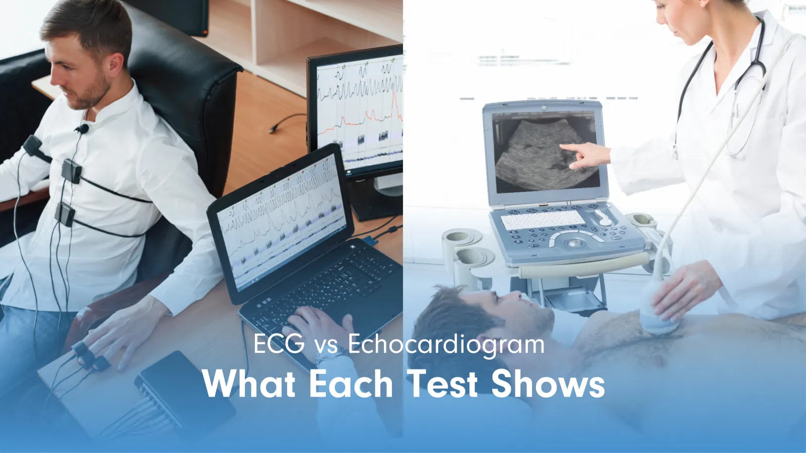
An ECG and an echocardiogram are both cardiac tests, but they measure different things. An ECG records the electrical activity of the heart. An echocardiogram produces images of the heart’s structure and movement using sound waves.
What an ECG Measures
An electrocardiogram (ECG) records the electrical impulses produced by the heart with each beat. RadiologyInfo.org’s ECG overview, published by the Radiological Society of North America, describes the ECG as a test that charts heart rhythm in the form of waves that reflect how electrical signals travel through the heart muscle. The pattern and timing of those waves tells the physician if the rhythm is normal, irregular, too fast, or too slow.
Electrodes are attached to the chest, arms, and legs. The procedure takes fewer than 10 minutes and requires no preparation. No radiation is used and the test is entirely painless.
What an ECG Can Detect
- Arrhythmias: Irregular, too-fast, or too-slow heart rhythms detected through abnormal wave patterns.
- Heart attack evidence: ST-segment changes in the waveform indicate current or prior myocardial infarction.
- Conduction abnormalities: Bundle branch blocks and other delays in electrical conduction through the heart muscle.
- Chamber enlargement: Certain wave patterns suggest enlarged heart chambers, which can indicate underlying conditions.
What an Echocardiogram Measures
An echocardiogram uses high-frequency sound waves to produce live images of the heart’s chambers, valves, walls, and major vessels. RadiologyInfo.org’s echocardiography page describes it as a tool that shows how well the heart is beating and pumping blood, and if structural abnormalities are present. A gel is applied to the chest and a transducer is moved across the skin to capture images, similar to an abdominal ultrasound.
What an Echocardiogram Can Detect
- Valve disease: Narrowing (stenosis) or leaking (regurgitation) of any of the four heart valves.
- Heart failure: Reduced ejection fraction, indicating the heart muscle is not pumping blood effectively.
- Wall motion abnormalities: Areas of the heart wall that are not contracting normally, often due to prior ischaemia.
- Pericardial effusion: Fluid accumulation around the heart that can compress cardiac function.
- Congenital defects: Structural abnormalities present from birth, including septal defects and valve malformations.
ECG vs Echocardiogram: Key Differences
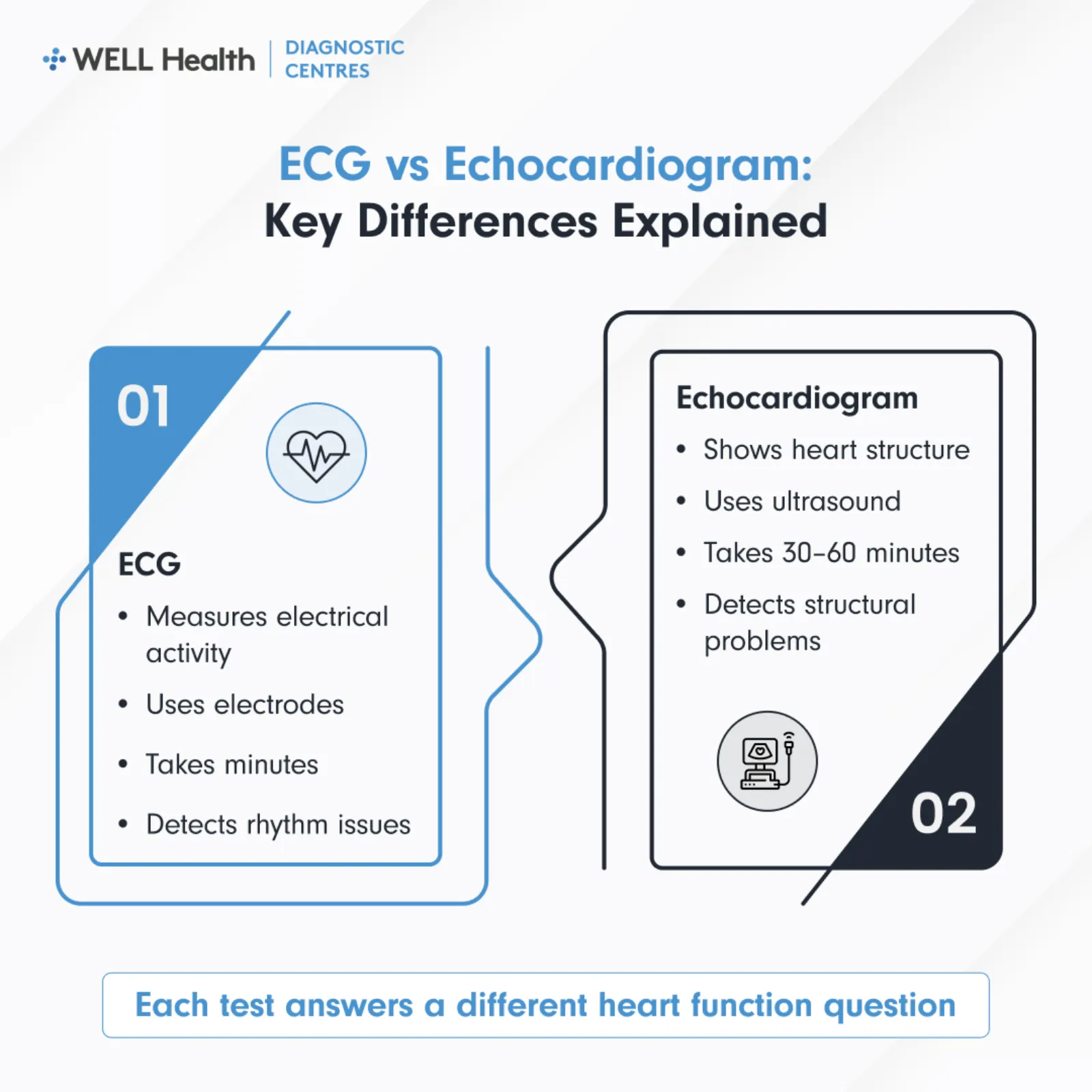
An ECG shows how the heart’s electrical system is working, while an echocardiogram shows how the heart looks and moves. Both tests are commonly used to evaluate heart health, but they answer different questions. Understanding these differences can help you know why one test may be ordered over the other, or why your doctor may recommend both.
Technology Used
An ECG uses electrodes placed on the skin to detect and record electrical signals. An echocardiogram uses a transducer and ultrasound technology to generate images. One measures electrical function; the other visualizes mechanical and structural function.
Duration and Preparation
An ECG is completed in under 10 minutes and requires no preparation. An echocardiogram typically takes 30 to 60 minutes. Neither test involves radiation, fasting, or any recovery period.
What Each Test Answers
An ECG answers questions about heart rhythm, rate, and conduction. An echocardiogram answers questions about heart structure, valve function, wall movement, and pumping efficiency. A physician may order both when they need a complete picture of both the electrical and structural aspects of the heart.
When Each Test Is Ordered
Physicians select the appropriate test based on the clinical question and the patient’s presenting symptoms.
When an ECG Is Ordered
- Palpitations or irregular heartbeat: The ECG identifies if the rhythm is clinically significant.
- Chest pain: An ECG is typically the first cardiac test in an acute chest pain assessment.
- Fainting or dizziness: Used to rule out electrical causes of loss of consciousness or lightheadedness.
- Pre-operative clearance: Many surgical protocols require a baseline ECG before anaesthesia.
- Routine cardiac monitoring: For patients with known heart disease, diabetes, or hypertension, on a regular review schedule.
When an Echocardiogram Is Ordered
- Shortness of breath: To assess if the heart’s pumping function or valve function is contributing to symptoms.
- Heart murmur: To identify the source, severity, and clinical significance of an abnormal sound on auscultation.
- Suspected heart failure: To measure ejection fraction and evaluate chamber size and wall motion.
- Atrial fibrillation: To assess for structural causes and check for blood clots in the heart before cardioversion.
- Follow-up after heart attack: To evaluate residual wall motion abnormalities and overall cardiac function.
When Additional Cardiac Tests Are Needed
An ECG or echocardiogram result may lead to further cardiac investigation depending on the findings. A Holter monitor extends ECG recording over 24 to 48 hours to capture intermittent arrhythmias that appear unpredictably. A stress echocardiogram combines exercise or pharmacological stress with echocardiography to assess how the heart performs under load.
Key Takeaways
- Different measurement types: An ECG captures electrical signals; an echocardiogram captures structural images.
- Different technology: An ECG uses skin electrodes; an echocardiogram uses ultrasound.
- Different appointment length: An ECG takes minutes; an echocardiogram takes 30 to 60 minutes.
- Ordered for different symptoms: ECG is ordered for rhythm concerns; echocardiogram is ordered for structural or functional concerns.
- Can be ordered together: Both tests are sometimes ordered at the same visit when a complete cardiac assessment is needed.
Frequently Asked Questions
Can an ECG detect the same things as an echocardiogram?
No. An ECG detects electrical abnormalities such as arrhythmias and conduction problems. An echocardiogram detects structural and functional abnormalities such as valve disease, wall motion issues, and reduced ejection fraction. The two tests complement each other and cannot substitute for one another in clinical assessment.
Which test is more commonly ordered first?
An ECG is typically the first cardiac test ordered in most clinical presentations because it is fast, non-invasive, and available in most clinical settings. An echocardiogram is ordered when the clinical question involves heart structure or function, which an ECG cannot assess. Both tests may be ordered at the same time when a complete cardiac workup is indicated.
Is an echocardiogram the same as a cardiac ultrasound?
Yes. An echocardiogram is a type of cardiac ultrasound. The terms are used interchangeably in clinical practice. It uses the same ultrasound technology as abdominal or obstetric imaging, applied specifically to assess heart structure and function.
ECG and Echocardiogram as Part of Cardiac Assessment
An ECG and echocardiogram each address a different dimension of cardiac function. Used together, they provide a comprehensive assessment of both the electrical and mechanical aspects of the heart. Your physician determines which test or combination of tests is appropriate based on your symptoms and clinical history.